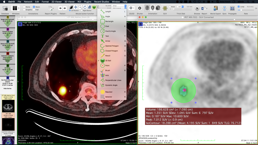
Quadrant I: Coronal reconstruction in the upper-right pane.jpg files for subsequent importation into Plaque Simulator. Export axial, coronal, sagittal, tumor-coronal and tumor-meridian reconstructions as.Adjust the three orthogonal views to align with the appropriate eye.Select the 3D Curved-MPR (Multi Planar Reconstruction) menu item.Click on the 2D/3D button at the top of the window.Double-click on the desired study: e.g.The loaded image sets will appear in the patient database viewer window of the application.

If it doesn't automatically find the images, open the CD (or portable device) from the MacOSX Finder and drag the folder containing the images onto the application window or its icon in the dock.

These 6 reconstructions will be imported into Plaque Simulator and used to measure the size and shape of the eye and, in conjunction with ultrasound images and fundus photos, to determine the tumor size, shape and location within the eye. The user interfaces are nearly identical.

Horos (son of Osiris) is derived from the same code base as OsiriX but is free to use. OsiriX predates Horos but is now a paid, FDA approved product. Other reconstructions of interest are the meridian and coronal planes which intersect the tumor apex. Horos and OsiriX are DICOM listeners and viewers that can manipulate 3-D data sets and export reconstructions of axial, equatorial and sagittal planes which bisect the affected eye as.


 0 kommentar(er)
